Cardiac Imaging: What It Is and Why It Matters
If your doctor mentioned a heart scan, you might wonder what actually happens inside the machine. Cardiac imaging is simply a group of tests that create pictures of your heart so doctors can see how well it’s pumping, spot blockages, or catch early signs of disease. The good news is most of these tests are quick, painless, and give incredibly detailed information that helps guide treatment.
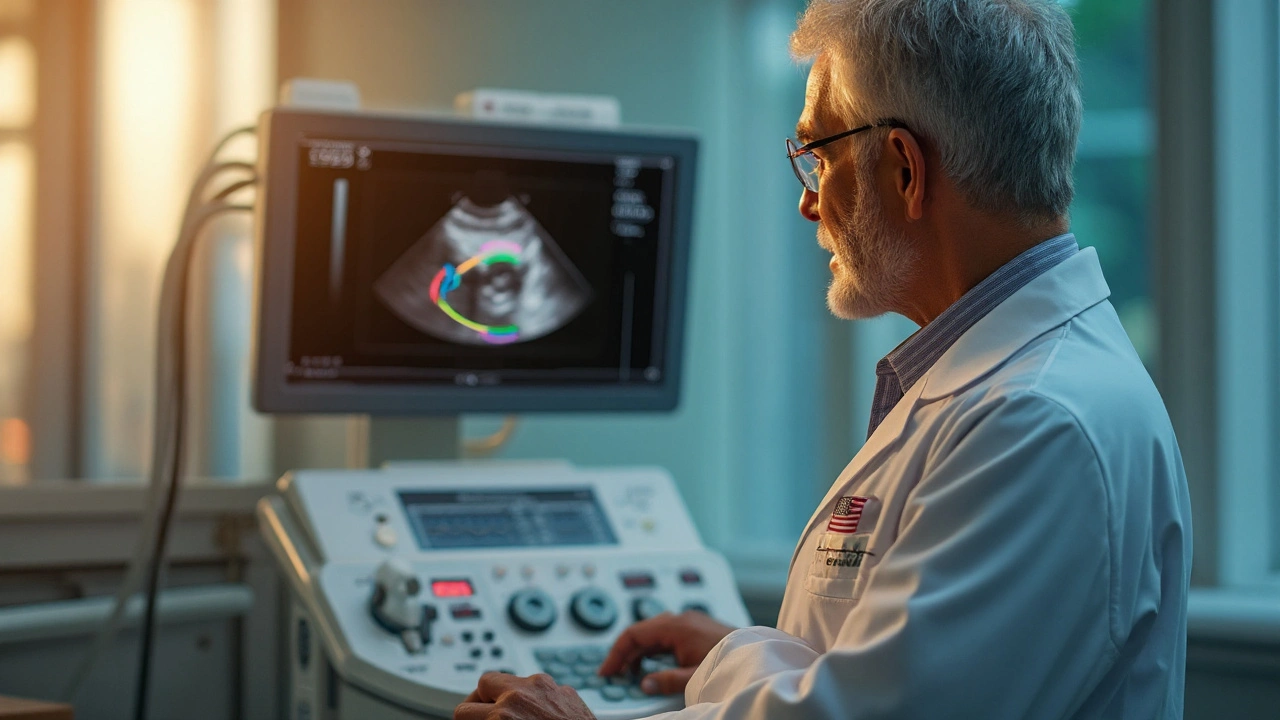
Common Cardiac Imaging Techniques
Echocardiogram (Echo) uses sound waves to produce moving images of the heart. It’s the most common first‑line test because it’s cheap, safe, and doesn’t involve radiation. The technician puts a gel‑filled probe on your chest and slides it around while you lie on a table. You’ll see the heart beating on a screen in real time, and the doctor can measure valve function, wall thickness, and blood flow.
Cardiac MRI takes pictures with magnetic fields and radio waves. It’s great for looking at heart muscle tissue, checking for scarring after a heart attack, or assessing congenital defects. You’ll lie in a narrow tube for about 30–45 minutes while the machine hums. Some people feel claustrophobic, but most centers offer open‑type scanners or a mirror to watch a movie.
Cardiac CT (Coronary CT Angiography) creates a 3‑D view of the coronary arteries using X‑rays. It’s often used to rule out blockages in low‑risk patients or to plan procedures like stent placement. You’ll receive a small injection of contrast dye, hold your breath for a few seconds, and the scanner spins around you. The radiation dose is low compared to older techniques, but it’s still higher than an echo.
Stress Tests with Imaging combine exercise or medication‑induced heart stress with either echo or nuclear imaging. The goal is to see how the heart performs under pressure. If blood flow drops in a region, it could indicate a narrowing that needs further attention.
Preparing for Your Heart Scan
Most cardiac imaging tests require minimal preparation, but a few tips can help the process go smoothly. For any test that uses contrast dye (CT or certain MRIs), drink plenty of water beforehand and inform the staff if you have kidney issues or allergies. If you’re scheduled for an echo, you can eat and drink normally.
When a stress test is ordered, avoid caffeine and nicotine for at least 12 hours, as they can affect heart rate and blood pressure readings. Wear comfortable clothing without metal zippers or buttons, especially for MRI, because metal can interfere with the magnetic field.
Ask the technician about what you’ll hear during the scan. The sounds are usually just the machine’s whirring or a series of clicks—nothing to worry about. If you’re anxious, let the staff know; they can offer a mild sedative or a calming environment.
After the scan, you can usually resume normal activities right away. If you received contrast dye, drink extra fluids to help flush it out. Your doctor will review the images and discuss results within a few days, either by phone or during a follow‑up visit.
Understanding the basics of cardiac imaging makes the whole experience less intimidating. Whether you’re getting an echo to check a murmur or a CT to rule out coronary disease, these tools give doctors a clear picture of your heart’s health, leading to faster and more precise care.
How Echocardiography Detects Left Ventricular Dysfunction - A Practical Guide
Learn how echocardiography identifies left ventricular dysfunction, the key measurements involved, when to use advanced imaging, and practical steps for clinicians.
Read more