How Echocardiography Detects Left Ventricular Dysfunction - A Practical Guide
 Sep, 25 2025
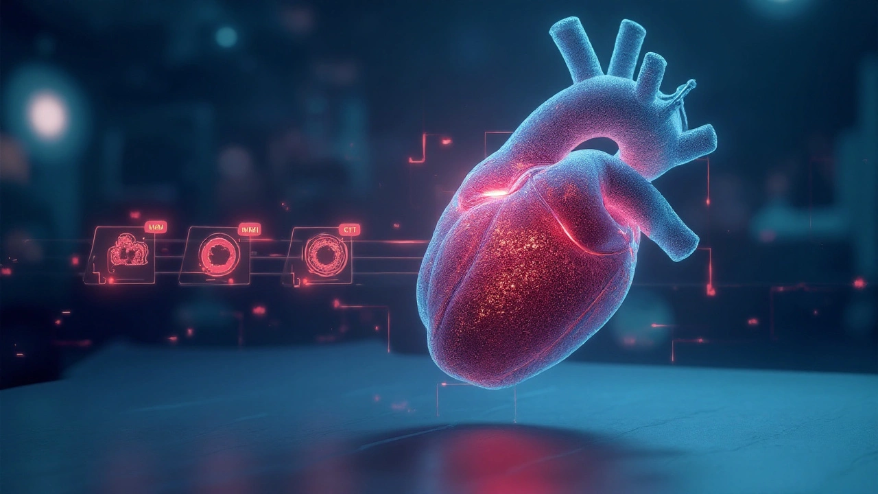
Echocardiography is a non‑invasive ultrasound technique that visualises heart structures in real time, characterised by its ability to quantify chamber size, wall motion and blood flow without ionising radiation. In everyday practice, it’s the first‑line tool for spotting left ventricular dysfunction, a condition that underpins most cases of heart failure.
What Is Left Ventricular Dysfunction?
Left ventricular (LV) dysfunction describes the heart’s main pumping chamber failing to contract or relax properly. It splits into two classic patterns:
- Systolic dysfunction - reduced ejection fraction (EF) below 50%; the ventricle can’t push blood out effectively.
- Diastolic dysfunction - preserved EF but stiff walls that hinder filling, leading to elevated pressures.
Both forms raise the risk of heart failure, arrhythmias and reduced exercise capacity. Early detection lets clinicians start guideline‑directed therapy before irreversible remodeling sets in.
How Echocardiography Works
The ultrasound transducer emits high‑frequency sound waves that bounce off cardiac tissues. The returning echoes are processed into moving images on a screen. Two core modes dominate LV assessment:
- 2D echocardiography - provides planar slices of the heart, ideal for measuring chamber dimensions and visualising wall motion.
- Doppler echocardiography - measures blood velocity, enabling calculation of pressure gradients and flow‑related indices.
Advanced add‑ons like tissue Doppler imaging (TDI) and speckle‑tracking strain imaging capture subtle myocardial mechanics that precede overt EF decline.
Key Echocardiographic Parameters for LV Dysfunction
When you open an echo study, the first numbers you look for are:
- Ejection fraction (EF) - percentage of blood expelled each beat; normal 55‑70%.
- LV end‑diastolic volume (EDV) - cavity size at filling; enlarged values hint at chronic overload.
- Global longitudinal strain (GLS) - percentage shortening of myocardial fibers; abnormal when <‑16%.
- E/e′ ratio - Doppler estimate of filling pressure; >14 suggests diastolic dysfunction.
Below is a quick comparison of the most common imaging tools used to assess LV function.
| Modality | Typical EF Accuracy | Ability to Detect Subtle Dysfunction | Availability in Community Settings |
|---|---|---|---|
| 2D Echocardiography | ±5% | Low (relies on visual EF) | High |
| Tissue Doppler Imaging | ±4% | Medium (assesses systolic & diastolic velocities) | Moderate |
| Speckle‑Tracking Strain | ±3% | High (GLS detects early fibre loss) | Increasing, but vendor‑specific |
| Cardiac MRI | ±2% | Very High (gold standard for volumes & strain) | Limited to tertiary centres |
Practical Workflow: From Referral to Report
Most hospitals follow a standard pathway:
- Referral - a cardiologist or GP suspects LV disease based on symptoms, ECG changes or elevated BNP levels.
- Acquisition - a trained sonographer obtains the standard views (parasternal long‑axis, short‑axis, apical 4‑chamber, etc.).
- Analysis - the interpreting cardiologist measures EF, volumes, GLS and Doppler indices, referencing the American Society of Echocardiography guidelines for normal cut‑offs.
- Report - concise narrative with quantitative values, severity grading (mild, moderate, severe) and recommendations for follow‑up or additional testing.
Key pitfalls to avoid: poor acoustic windows, off‑axis cuts, and forgetting to optimise gain settings - all can skew EF calculations.
When Echo Falls Short
Although echo is versatile, it has limits:
- Obesity or lung disease may produce sub‑optimal images.
- EF can appear normal in early diastolic dysfunction, hiding high filling pressures.
- Quantifying scar tissue or complex congenital anatomy often requires Cardiac MRI, which offers superior tissue characterization.
In such cases, clinicians supplement echo with cardiac MRI, stress testing or invasive haemodynamics.
Emerging Techniques and Future Directions
Three trends are reshaping LV assessment:
- 3‑D volumetric echo - captures the entire ventricle in one dataset, reducing geometric assumptions.
- Contrast‑enhanced echo - improves border delineation in low‑quality windows, boosting EF accuracy.
- Artificial‑intelligence algorithms - automate EF and strain calculations, cutting inter‑observer variability.
Early adoption studies show AI‑derived EF agrees within ±2% of expert measurements, promising faster turn‑around in busy clinics.
Take‑Home Checklist for Clinicians
- Confirm the indication: symptoms, ECG, BNP, or routine screening.
- Obtain high‑quality 2D images and Doppler tracings.
- Measure EF, LV volumes and GLS; document any wall‑motion abnormalities.
- Cross‑check Doppler‑derived filling pressures (E/e′) for diastolic assessment.
- If images are poor or suspicion remains high, consider cardiac MRI or contrast echo.
By following these steps, you’ll catch LV dysfunction early, start evidence‑based therapy, and improve patient outcomes.
Frequently Asked Questions
What is the normal range for left ventricular ejection fraction?
A healthy adult typically has an EF between 55% and 70%. Values below 50% suggest systolic dysfunction, while 50‑55% may be borderline and warrant closer follow‑up.
How does speckle‑tracking strain improve early detection?
Strain measures the percentage shortening of myocardial fibres. Global longitudinal strain (GLS) often becomes abnormal (<‑16%) before EF drops, identifying patients at risk even when conventional echo looks normal.
When should I order cardiac MRI instead of echo?
Consider MRI when echo windows are poor, when precise quantification of volumes or scar tissue is needed (e.g., after myocardial infarction), or when you need tissue characterization for infiltrative diseases like amyloidosis.
Can echocardiography detect diastolic dysfunction reliably?
Yes, by using Doppler measurements such as the E/e′ ratio, left atrial volume, and pulmonary vein flow patterns. Combining these indices with clinical data provides a robust grading of diastolic impairment.
What are the common pitfalls that lead to inaccurate EF measurements?
Mistakes include off‑axis apical views, inadequate end‑systolic frame selection, neglecting to adjust for foreshortening, and using poor image quality. Using biplane Simpson’s method and verifying trace accuracy reduce these errors.
Daisy Aguirre
September 25, 2025 AT 06:12What a vivid tour through the echo world! Your guide spins a rainbow of terminology that even a non‑cardiologist can chase. I love how you broke down EF, GLS and the newer 3‑D tricks into bite‑size nuggets. The checklist at the end feels like a friendly coach shouting, “You’ve got this!” Keep sprinkling those colorful analogies – they make the heavy tech feel breezy.
Natalie Kelly
October 6, 2025 AT 19:59Great summary, lol. I love the step‑by‑step checklist.
Tiffany Clarke
October 18, 2025 AT 09:45Echo is the Swiss Army knife of cardiac imaging.
Sandy Gold
October 29, 2025 AT 22:32While the article covers the basics, it glosses over the nuances of strain analysis. One must recognize that GLS is not a universal surrogate for contractility across all vendor platforms. Moreover, mentioning “AI‑derived EF” without discussing algorithmic bias feels like marketing fluff. The table could have benefited from confidence intervals rather than single point estimates. Finally, I’d have liked a deeper dive into diastolic parameters beyond just E/e’.
Frank Pennetti
November 10, 2025 AT 12:19This guide is a textbook rehash; nothing new for seasoned cardiologists.
Adam Baxter
November 20, 2025 AT 22:19Love the energy here! Keep pushing those AI frontiers. The future is bright for bedside echo.
Keri Henderson
November 30, 2025 AT 04:32Super helpful steps for newbies. Make sure to practice the apical 4‑chamber view until it feels natural. Consistency beats perfection every time.
elvin casimir
December 8, 2025 AT 20:52Interesting take, but there are a few slip‑ups: you wrote “GLS detects early fibre loss” – it’s actually “myocardial fibre shortening”. Also, “AI‑derived EF” should be qualified as “algorithm‑estimated EF”. Precision matters when trainees copy‑paste.
Steve Batancs
December 15, 2025 AT 19:32I appreciate the comprehensive overview and the clear structure of the workflow. The inclusion of both 2D and Doppler modalities reflects best practice guidelines.
Ragha Vema
December 21, 2025 AT 14:25Reading this feels like stepping onto a stage where the spotlight blazes on every echo machine in the world. First, the author paints 2D echo as the humble workhorse, but we all know the hidden agenda-manufacturers pushing upgrades like the latest AI filter that promises "clinical perfection". Second, the mention of speckle‑tracking is almost sacramental, yet the reality is that many community hospitals lack the software licenses, leaving clinicians stranded with outdated numbers. Third, the table comparing modalities reeks of selective bias; why omit cost and training time? Fourth, the checklist sounds reassuring, but it ignores the fact that poor acoustic windows can masquerade as normal EF, leading to false reassurance. Fifth, the AI hype is unsettling-who audits the black‑box decisions, and what happens when the algorithm misclassifies a borderline case? Sixth, while contrast‑enhanced echo is praised, the risks of microbubble reactions are barely mentioned. Seventh, the “early adoption studies” claim ±2% agreement, yet those studies were sponsored by the same vendors pushing the tech. Eighth, the guide fails to address the ethical dilemma of over‑diagnosing subclinical dysfunction, which can lead to unnecessary medication. Ninth, I recall a case where a strained GLS figure led to a premature ICD implantation-an outcome nobody wanted. Tenth, the absence of patient‑centered outcomes, like quality‑of‑life measures, makes the whole narrative feel mechanistic. Eleventh, the article skips the reality of insurance reimbursement hurdles that keep advanced echo tools out of reach for many practices. Twelfth, there is no discussion of operator fatigue, which can subtly degrade image quality over a long shift. Thirteenth, the reference to “guideline‑directed therapy” assumes all clinicians have equal access to the latest trials. Fourteenth, the ever‑present specter of data privacy looms when AI systems store imaging datasets on cloud servers. Fifteenth, and perhaps most importantly, the piece glosses over the human element: the skill, intuition, and bedside rapport that no machine can replicate. In short, while the technology dazzles, we must keep our feet on the ground and remember that echo is a tool, not a replacement for thoughtful clinical judgment.
Scott Mcquain
December 26, 2025 AT 05:32Indeed, the points raised are, without doubt, compelling; however, one must, perhaps, temper the tone with a measured acknowledgment of the undeniable advancements that have, undeniably, transformed clinical practice, and, consequently, patient outcomes.
kuldeep singh sandhu
December 29, 2025 AT 16:52While the critique is thorough, I think the emphasis on AI overshadows the simple beauty of a well‑performed manual echo.
Mariah Dietzler
January 1, 2026 AT 00:25Seems okay, but could use more images.
Nicola Strand
January 2, 2026 AT 04:12Although the article is comprehensive, it neglects the cost‑effectiveness of handheld devices, which are increasingly vital in low‑resource settings.