What Is Echocardiography and Why It Matters
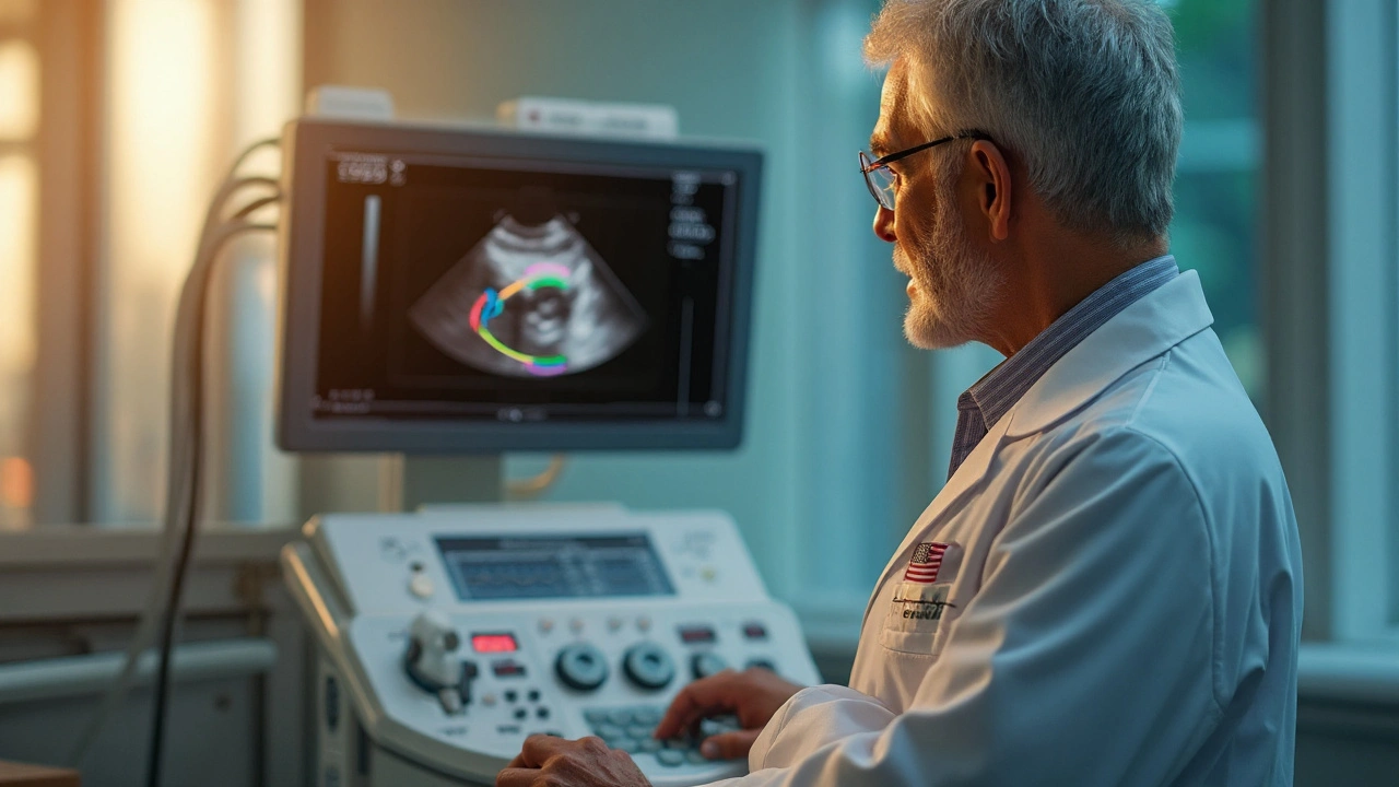
Echocardiography, or simply an echo, is a painless ultrasound of your heart. It lets doctors watch the chambers, valves and blood flow in real time, without any radiation. Think of it as a moving picture of your ticker, helping spot problems early before they become serious.
Types of Echo Tests You Might Hear About
The most common is the transthoracic echo (TTE). A technician glides a small probe across your chest, sends sound waves into the heart, and captures images on a screen. If a clearer view is needed—like checking a leaky valve or a clot—a transesophageal echo (TEE) is done. Here the probe goes down the throat, giving a closer look at the back of the heart.
Stress echo is another variant. You either jog on a treadmill or receive medication that makes the heart work harder, then the echo shows how well it handles the extra load. Doctors love it for spotting hidden coronary artery disease.
When and Why Your Doctor May Order an Echo
Typical reasons include unexplained shortness of breath, chest pain, irregular heartbeats, or a heart murmur heard during a physical exam. If you’ve had a heart attack, heart failure, or valve surgery, an echo tracks how well the heart is healing. It’s also the go‑to test for evaluating congenital heart defects in both kids and adults.
Because it’s quick and non‑invasive, an echo often replaces more expensive or risky procedures. A normal result can spare you from unnecessary medication, while an abnormal finding guides the right treatment plan.
What to Expect During the Appointment
Arrive in comfortable clothing—short sleeves are best for TTE. The tech will apply a warm gel on your chest; it helps the probe glide and improves the sound waves. The scan usually lasts 30‑45 minutes. You’ll lie still while the images are captured, and the tech may ask you to hold your breath briefly to get clearer pictures.
If you’re having a TEE, you’ll be given a mild sedative, and a throat spray numbs the area. The probe feels like a soft tube sliding down your throat, but most people tolerate it well. Recovery is quick; you’ll be cleared to eat once the numbness wears off.
Understanding the Results
The echo report lists measurements such as ejection fraction (how much blood the left ventricle pumps each beat), valve thickness, and any regurgitation (leakage). A normal ejection fraction is 55‑70 %. If your numbers are lower, your doctor might suggest medication, lifestyle changes, or further testing.
Sometimes the report includes video clips of specific heart motions. Your physician can walk you through these, pointing out what looks healthy and what needs attention. Don’t hesitate to ask for plain‑language explanations—knowing the “why” helps you follow treatment plans.
Safety and Comfort Tips
Echoes are safe for everyone, including pregnant women and kids, because they use no ionizing radiation. If you have a pacemaker or other implanted device, let the tech know; the probe can be positioned to avoid interference.
Stay hydrated before a TTE, and avoid heavy meals prior to a stress echo. For a TEE, follow any fasting instructions your doctor gives. After the test, drink water to flush out the gel and ease any lingering throat soreness.
Quick FAQ
Do I need a referral? Most clinics require a doctor’s order, but some centers allow direct scheduling for routine checks.
Can I drive after a TEE? Yes, once the sedative wears off—usually within an hour. Have a friend ride along if you feel groggy.
How often should I get an echo? That depends on your heart condition. Some people need annual checks; others only when symptoms change.
Bottom line: an echocardiogram gives a clear, real‑time snapshot of your heart’s health without pain, radiation, or long waits. If your doctor suggests one, you’re getting a powerful tool to guide the right care.
How Echocardiography Detects Left Ventricular Dysfunction - A Practical Guide
Learn how echocardiography identifies left ventricular dysfunction, the key measurements involved, when to use advanced imaging, and practical steps for clinicians.
Read more